Medical imaging stands at the forefront of modern healthcare diagnostics, enabling doctors to peer inside the human body without making a single incision. Among the many imaging techniques available today, X-rays and CT scans (Computed Tomography) are two of the most frequently used tools. Both play crucial roles in helping healthcare professionals diagnose and monitor a variety of conditions, but they operate in remarkably different ways and serve unique purposes. For anyone navigating healthcare decisions—whether you're a patient, caregiver, or medical enthusiast—it’s invaluable to grasp the distinctions between these modalities. Understanding how X-rays and CT scans differ will help you appreciate their uses, benefits, and potential risks, ultimately enabling more informed discussions with your healthcare team.
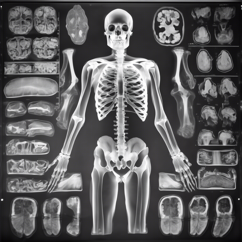
X-rays harness the power of high-energy electromagnetic radiation to create images of the inside of the body, particularly excelling at showing dense tissues like bones. When you undergo an X-ray exam, a small amount of radiation penetrates your body, while an external detector captures the pattern of rays absorbed by different tissues, forming a two-dimensional image. This imaging method has been around for over a century—credit goes to Wilhelm Conrad Roentgen, who discovered X-rays back in 1895, an event that revolutionized medicine almost overnight. Since then, X-rays have been widely used to diagnose broken bones, pinpoint infections, and detect certain kinds of cancers. Their speed, affordability, and simplicity make them a favorite first step in many diagnostic processes. Fun fact: the iconic “bones” images seen in many Halloween decorations actually stem from the eerie, ghostly shadows cast by early X-ray images!
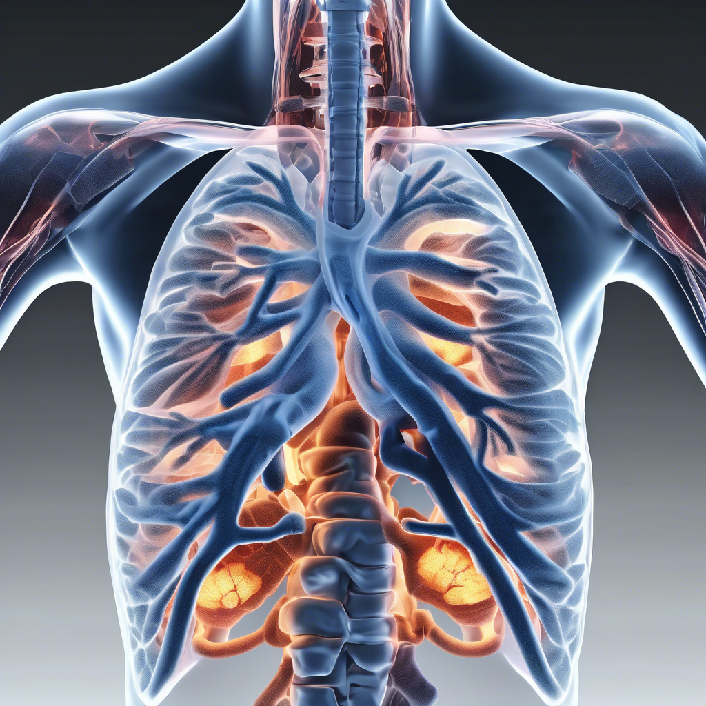
In contrast, CT scans take medical imaging a giant leap forward by amalgamating multiple X-ray images captured from several angles around the body, which are then processed by sophisticated computers to generate detailed cross-sectional and three-dimensional images. This technology, sometimes called a CAT scan, permits doctors to look at bones, muscles, fat, and organs with greater clarity and depth. CT scanners rotate around the patient, capturing numerous slices or “sections” that reveal intricate internal pictures, helping detect complex fractures, tumors, infections, and vascular diseases that would be challenging to diagnose with regular X-rays. While a CT scan takes longer and is generally more expensive than a standard X-ray, the level of detail it provides is often indispensable for a comprehensive diagnosis and treatment planning. Interestingly, the first full-body CT scan was performed in the early 1970s, and this breakthrough was recognized by the Nobel Prize in Physiology or Medicine awarded to Godfrey Hounsfield and Allan Cormack in 1979.
One of the key considerations when comparing X-rays and CT scans is radiation exposure. Both utilize ionizing radiation, which, while powerful for imaging, carries inherent health risks, particularly with repeated or high-dose exposure. Standard X-rays involve a relatively low dose of radiation and are generally considered safe for routine use. However, CT scans expose patients to significantly higher doses due to the multiple X-ray images required to construct detailed views from various angles. This difference means that CT scans are generally reserved for situations where the detailed visualization is crucial in diagnosis and treatment decisions, outweighing the slightly increased risk. Modern CT machines, though, are evolving swiftly; many incorporate dose-reduction technologies designed to minimize radiation exposure without compromising image quality. In the broader landscape, alternative imaging modalities such as MRI and ultrasound—which don’t involve ionizing radiation—are recommended whenever appropriate, offering safer long-term options for patients.
Technological advancements in medical imaging continue to push boundaries, marrying enhanced diagnostic clarity with patient safety. Cutting-edge CT scanners now employ iterative reconstruction algorithms and optimized scanning protocols that reduce radiation doses dramatically compared to early machines. Additionally, hybrid imaging techniques are emerging, combining modalities like PET-CT or PET-MRI that synergize the strengths of multiple methods to improve diagnoses, especially in oncology and neurology. Beyond the machines themselves, innovations like OncoPreventer highlight the power of integrating medical imaging with personalized health management tools. OncoPreventer offers tailored cancer screening plans, timely alerts for check-ups, and evidence-based guidance, empowering individuals to participate actively in preventing and detecting cancer early. Such proactive approaches complement the diagnostic prowess of medical imaging, illustrating how technology and personalized medicine herald a new era in healthcare.
In conclusion, while X-rays and CT scans both play indispensable roles in medical diagnostics, their applications, advantages, and limitations differ considerably. X-rays are best suited for quick assessments of bone injuries and some lung conditions, offering a low-cost, rapid diagnostic tool. CT scans provide a more detailed and three-dimensional insight, indispensable for investigating complex fractures, tumors, infections, and vascular abnormalities. Awareness of their differences, especially related to radiation exposure and diagnostic capabilities, equips patients and healthcare providers to select the most appropriate imaging strategy for each clinical scenario. Coupling these diagnostic tools with forward-thinking health management solutions like OncoPreventer promises a future where early detection, effective treatment, and patient empowerment walk hand in hand towards better health outcomes.
#MedicalImaging #XrayVsCT #HealthTech #Radiology #OncoPreventer #HealthcareInnovation #EarlyDetection