The integration of extended reality (XR) technologies—encompassing virtual reality (VR), augmented reality (AR), and mixed reality (MR)—has revolutionized surgical procedures by enhancing three-dimensional imaging techniques. Notably, MR blends digital overlays seamlessly with real-world visualizations, offering surgeons an unprecedented ability to interact with both physical and virtual elements in real time. This convergence of the virtual and physical realms enables a highly immersive and precise surgical experience, fundamentally transforming how complex surgeries are planned and executed.
Although MR’s applications have been predominantly explored in adult patients, especially in disciplines like cardiac surgery, neurosurgery, maxillofacial surgery, urology, orthopedics, and surgical oncology, its use in pediatric surgery remains relatively underrepresented, particularly in pediatric oncological cases. This emerging area is gaining momentum as researchers investigate how MR can address the unique challenges presented by pediatric patients, whose developing anatomical structures require meticulous surgical planning to preserve healthy tissue and ensure optimal long-term outcomes. Despite the scarcity of extensive studies focusing on pediatric surgical oncology, recent efforts have begun applying MR technology across a range of procedures—from thoracotomies and pulmonary metastasis resections to biopsies of femoral tumors, clavicle tumor resections, and resection of sacrococcygeal masses—illuminating MR’s versatility and potential in this delicate field.
One of the groundbreaking aspects of recent research involves not just the use of MR for visualization but also its dynamic interaction with the surgical environment. By generating 3D holograms from preoperative imaging studies like CT or MRI, surgeons can overlay these virtual models onto the patient’s body during surgery. This overlay is interactive, controlled via hand gestures, voice commands, and virtual menus, creating an intuitive interface that aids intraoperative navigation. For example, surgeons first align key anatomical landmarks and radiological markers between the hologram and the patient, then proceed with tumor resection while regularly referencing the hologram. This structured workflow—starting with preoperative planning, followed by intraoperative hologram application and guided resection—has been shown to improve surgical accuracy without prolonging operation times or increasing hospital stays. The educational benefits are significant as well; surgical teams, including experienced senior surgeons, report enhanced anatomical orientation and decision-making, underscoring the value of MR as both a planning and teaching tool in complex pediatric surgeries.
The promise of MR technology is accentuated when compared with traditional imaging methods. Studies consistently demonstrate that MR and 3D visualization, sometimes complemented by 3D printing, outperform conventional DICOM imaging in clarity, immediacy, and spatial understanding. For instance, in pediatric cardiac surgery, MR has been pivotal for preoperative planning, as shown by researchers like Gehrsitz and Brun. Others, like Chaussy, emphasize that 3D models allow surgeons to better anticipate procedural risks and select candidates for nephron-sparing surgeries. Furthermore, MR’s ability to localize pulmonary nodules with remarkable accuracy—achieving 94% precision versus 30% in manual palpation—highlights its potential to refine surgical navigation substantially. Importantly, MR-assisted procedures also display improved metrics such as reduced operative times, enhanced resection margins, decreased detection times in sentinel lymph node biopsies, and shorter task times in spatially complex liver surgeries. These cumulative benefits illustrate why experts advocate for broader adoption of MR systems in surgical oncology and beyond.
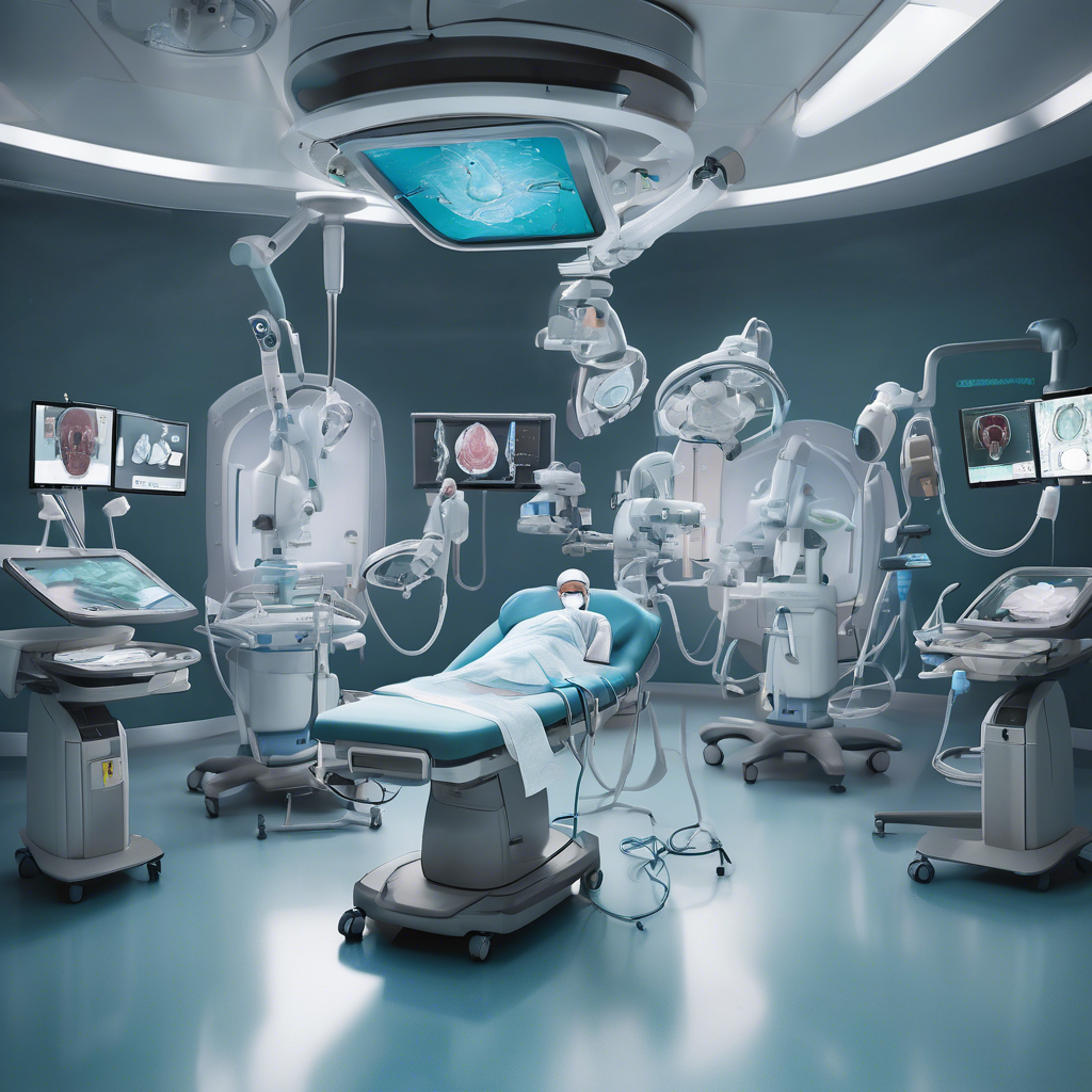
The CarnaLife Holo system exemplifies cutting-edge MR technology designed to elevate surgical workflow. Developed by MedApp S.A. in Poland, this platform converts medical imaging data into interactive holograms that surgeons can manipulate in three-dimensional space. Its proprietary software allows for detailed segmentation of tumors and surrounding anatomy, real-size overlays directly on patients, and intraoperative measurements without breaching sterile fields. The system features a high-performance workstation connected wirelessly to a Microsoft HoloLens 2 head-mounted display (HMD), facilitating real-time interaction through voice commands and hand gestures. Prior to surgery, imaging data in DICOM format are uploaded, with users enhancing visualization via adjustable presets and segmentation tools. During operations, radiological markers and anatomical landmarks guide hologram positioning, while printed reference points in the operating room ensure stable alignment even amid patient movements. Surgeons can visualize internal anatomy from multiple angles by manipulating the hologram with head movements, aiding in precise tumor localization and navigational accuracy. This integration of high-fidelity imaging and interactive holography heralds a new era where surgeons operate with augmented insight, blending technology and skilled craftsmanship.
Despite its transformative potential, MR technology faces several challenges. Real-time alignment of holograms with dynamic anatomical structures, especially when tissues deform during surgery, remains technically complex. Ergonomic considerations such as the weight and design of HMDs can impact surgeon comfort, causing fatigue or neck strain over prolonged procedures. Visual limitations including restricted field of view and parallax effects—where background and foreground elements shift at different speeds—may also affect usability. Battery life constraints of headsets impose additional operational hurdles during extensive surgeries. Economic factors cannot be overlooked: high initial costs for hardware, software development, and training could limit widespread adoption without thorough cost-benefit analyses. Encouragingly, preliminary findings suggest MR’s ability to reduce operative durations and hospital stays may offset these expenses through improved resource utilization and patient outcomes. As technology evolves, integrating MR with complementary modalities such as robotics, ultrasound, and artificial intelligence could further streamline workflows and enable dynamic adjustments during surgeries, helping overcome current limitations.
Looking forward, the future of MR in pediatric oncological surgery is bright and promising. Beyond pure visualization, incorporating factors like real-time tissue changes, AI-driven margin delineation, and integration with emerging robotic platforms could revolutionize surgical accuracy and safety. MR’s educational potential is equally exciting: holographic simulations provide immersive training environments where surgeons—both experienced and novice—can hone skills and anticipate complex scenarios. Furthermore, MR offers a pathway to better patient engagement, demystifying complicated procedures through vivid, interactive models. However, to translate this potential into routine practice, the surgical community must expand research with larger patient cohorts, apply objective measures to evaluate technological efficacy, and establish standardized protocols. Only then can MR transition from an innovative adjunct to a foundational component of pediatric oncological surgery, changing lives with precision and care.
#MixedReality #PediatricSurgery #SurgicalInnovation #MedicalTechnology #3DImaging #OncologicalSurgery #CarnaLifeHolo
Leave a Reply